Prior to carrying out the augmentation procedure, it is necessary to interview and examine the patient in compliance with the principles of medical art. Additional tests should include CBCT scanning or calibrated pantomographic x-ray. If there are any symptoms of chronic pathological changes to the nose or sinuses, a laryngological consultation should be ordered.
A CBCT scan or a calibrated pantomographic x-ray should be used to determine the height of residual bone or the height of the alveolar ridge at the location of the procedure. The width of the alveolar ridge should exceed the implant’s diameter by more than 3-4 mm. The width of the ridge can be increased during the surgery with the use of osteotome techniques. A precise assessment of the height of the bone is indispensable for a successful procedure.
- Drill P (pilot drill) is designated for marking the drilling position and performing a preliminary bone preparation to a desired depth.
- Drill P is designed to work exclusively with line>3< stoppers.
- The selection of the stopper should be based on the height of the residual bone assessed during a a radiological examination.

EXAMPLE:
If the height of the residual is 6 mm, the stopper to be fixed on the bur is marked as S3/6. The recommended speed for drill P is approx. 600 rpm with intensive external cooling. After making a preparation, it is necessary to assess the floor of the cavity with a calibrated probe from the implantation kit. The floor should be hard.
Next, the bone opening needs to be enlarged with a twist drill.
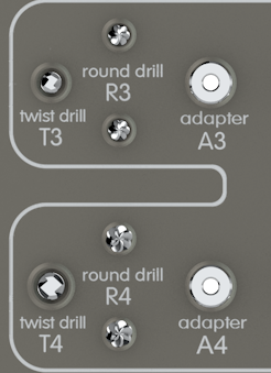
- When the diameter of the designed implant is approx. 4 mm, we switch to drill T3.
- When the diameter of the designed implant reaches approx. 5 mm, we apply drill T3 and then drill T4. The rotating speed for T3 and T4 is approx.. 600 rpm with intensive external cooling.
The drilling should be carried out by applying a minimum pressure up to the point of resistance.
In the case of poor bone quality or a narrow alveolar ridge, the bone bed may be enlarged by using appropriate osteotomes instead of drills T3 and T4. This results in the thickening of the bone structure and the crosswise expansion of the alveolar ridge. Osteotomes should be inserted carefully, without using a surgical mallet, up to the point of resistance.
Based on the diameter of the designed implant, it is then necessary to select an appropriate adaptor – A3 (implants with a diameter of approx. 4 mm) or A4 (implants with a diameter of approx. 5 mm), and fit it with a dedicated standard drill R3 or R4. The cutting part of drill R3 or R4 should adhere to the upper part of adaptor A3 or A4.
Using the hexagonal end of the hand wrench, it is then necessary to tighten two blocking screws located in the lower part of adaptor A3 or A4.
After that, a preset type torque wrench is placed on the hand wrench and, after selecting the torque moment of 15 Ncm, the blocking screws are tightened.
Now it is necessary to select an appropriate stopper compatible with the height of the alveolar ridge assessed during a radiological examination and fix it onto the adaptor with a fitted drill. Adaptor A3 functions with line >3< series stoppers, while adaptor A4 functions with line >4< series stoppers.
IMPORTANT!
The second digit in the stopper’s number always corresponds to the height of the residual bone given in millimeters.
The flat part of the hand wrench is used to loosen the stoppers if they get stuck on the adaptor.
The infini-Ti® sinus grafting kit includes additional elements described as “blocking screws” to be used for a misplaced blocking screw.
Tightening the blocking screw onto adaptors A3, A4, A5/2.8 and A5/4 is carried out through an opening located on the opposite side of the opening the blocking screw and with the use of the hexagonal part of the hand wrench.

IMPORTANT!
While dismantling drill R3 or R4, it is always necessary to loosen the blocking screws up to the point of resistance. Such procedure limits a possibility of spontaneous loosening of the blocking screw.
The recommended drilling speed of the established drilling system is 60-600 rpm.
Speeds exceeding 60 rpm require extensive external cooling, as well as applying “pumping” movements of the drill. The drilling should continue until the stopper rests on the alveolar ridge bone.
After completing the opening, it is necessary to use a calibrated, blunt-ended probe to check whether the mucous membrane of the sinus can be detected in the floor of the cavity.
IMPORTANT!
The probe should not penetrate the submucous area deeper than 1 mm above the floor of the maxillary sinus.
If the bone can still be detected, it is necessary to select a stopper 1 mm longer and repeat the procedure.
The integrity of the mucous membrane should be additionally verified by occluding the patient’s nostrils and asking the patient to blow the air gently through the nostrils (“nose blowing test”, Valsalva maneuver with the mouth open).
The next stage involves separating the mucous membrane of the cavity from bone edges with the use of micro elevators (the recommended type is the “mushroom elevator” with a mushroom shaped working tip).
The space prepared in the above way must now be filled with a biomaterial by using standard applicators (pluggers, syringes, osteotomes) and lightly condensed to make room for subsequent portions. The tools used to condense the biomaterial should be smaller in diameter than the prepared bone bed.
The biomaterial must be inserted very slowly.
IMPORTANT!
The tools used to condense the biomaterial should not be inserted deeper than the height of the residual bone.
After depositing an appropriate amount of biomaterial (0.1 ccm usually raises the mucous membrane by 1 mm) into the bone loss, the biomaterial should be gently moved in the applied direction, as well as sideways in order to create space necessary to insert the implant. It is recommended that the “nose blowing test”/Valsalva maneuver with the mouth open is repeated.
The procedure is completed by applying the implant and suturing the tissues . The selected implant should feature parameters providing for good mechanical stability.
The surgery should be followed up by a radiological examination.
Detailed description of the entire procedure is also available in a PDF file.
